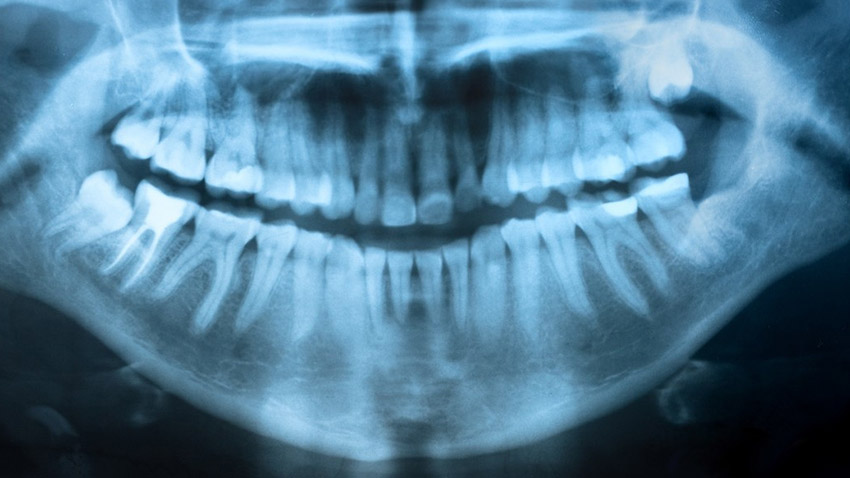
Dental X-Ray
Also known as dental radiographs, dental X-rays use controlled pulses of radiation to create images of the internal structures of the jaw and mouth.
How are dental X-rays taken?
Dental x-rays are taken with you sitting upright in a chair.
The dental technician will place a lead apron over your chest and wrap a thyroid collar around your neck. The x-ray sensor or film will be placed in your mouth for the picture. In the past, yearly x-rays were often recommended by dentists. But today, the ADA recommends that healthy adults with no major apparent dental problems only need to get x-rays about every 2-3 years. If your mouth is healthy and free of issues like gum disease and tooth decay.